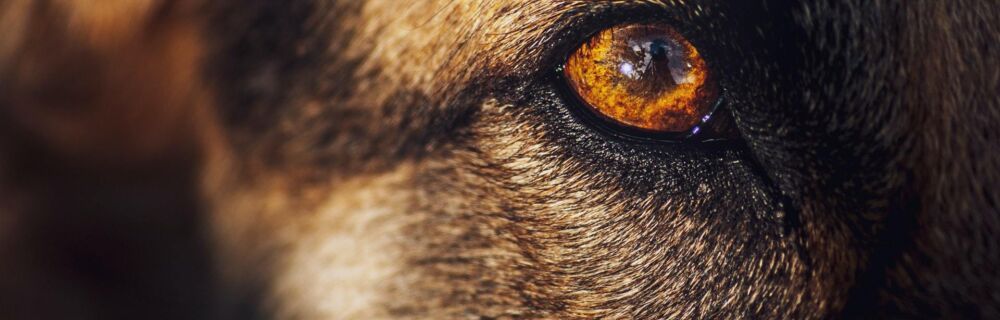
Why do my dog’s eyes look cloudy?
The cornea is the transparent protective layer covering the front surface of the eyeball. It helps to focus light as it enters the iris and the pupil. It also connects with the sclera (the white part of the eye) and serves as a barrier against debris and germs. Corneal changes, such as degeneration and dystrophy, are characterized by lipid or calcium deposits within the layers of the cornea. These appear as white or opaque areas on the surface of the eyeball, and rarely cause blindness, despite being very noticeable. Keep reading to learn more about the causes, diagnosis, and treatment for corneal degeneration and dystrophy in dogs.
Are you concerned about your pet?
Book a video consultation with an experienced veterinarian within minutes.
- Professional vet advice online
- Low-cost video vet consultations
- Open 24 hours a day, 365 days a year
Corneal Changes in Dogs
Corneal dystrophy affects both eyes. It can occur in dogs of any age and is an inherited trait in Airedales, Beagles, Cocker Spaniels, Miniature Schnauzers, Shelties, and Siberian Huskies.
Corneal degeneration is not inherited and can occur in one or both eyes. It frequently develops in areas of the cornea that have experienced trauma or chronic disease. In severe cases, the lipid or calcium deposits from corneal dystrophy and degeneration can cause discomfort from the roughened corneal surface, corneal ulceration, and ocular infections. In rare cases, dogs with corneal dystrophy/degeneration may have severe visual impairment or even blindness.
What causes these changes to a dog’s eye?
One of the most common causes of corneal degeneration are lipid deposits in the corneal stroma. These can be the result of increased cholesterol and triglycerides in the blood plasma.
Elevated cholesterol and triglycerides are frequently seen with endocrine diseases such as hypothyroidism, diabetes mellitus, and Cushing's disease.
Calcium deposits, which can be caused by elevated calcium levels in the blood, can also result in corneal degeneration, although they are less common than lipid deposits. Other disorders that can lead to an increased likelihood of corneal degeneration are pancreatitis, liver disease, hypophosphatemia (low phosphorus), and hypervitaminosis D.
Symptoms of Corneal Changes in Dogs
- White opacities in the cornea
- Roughened corneal surface
- Distinct margin between the cornea and the sclera
How are corneal dystrophy and degeneration diagnosed in dogs?
Diseases of the cornea are diagnosed with a fluorescein stain test. A drop of orange dye is placed into each eye and then the eyeball is visualized under a black light. If there is a defect in the surface of the cornea such as a corneal ulcer or scratch, the stain will gather in the defect and be highlighted by the black light. Although corneal degeneration and dystrophy can result in corneal ulceration, there are various other causes for ulcers on the surface of the eyeball. Corneal ulceration, if seen, will need to be treated before a diagnosis of corneal degeneration or dystrophy can be made.
If one or both eyes has a hazy, opaque appearance and the corneas do not retain fluorescein stain, corneal degeneration or dystrophy can be diagnosed. If the defect is in both eyes and the dog is an Airedale, Beagle, Cocker Spaniel, Miniature Schnauzer, Sheltie, or Siberian Husky, this is consistent with a diagnosis of corneal dystrophy. If the defect is only in one eye or if the dog is not one of the aforementioned breeds, this is considered corneal degeneration.
Treatment Options for Corneal Changes in Dogs
Mild cases of corneal degeneration and dystrophy can be treated with a topical acid treatment (TCA), which helps to dissolve the material. This treatment may need to be performed one or more times and is usually done under the care of a veterinary ophthalmologist. In severe cases, especially when there is significant discomfort, a surgical procedure to remove the outer layer of the cornea, called superficial keratectomy, is recommended. Corneal ulcers that result from corneal degeneration or dystrophy will need to be treated before considering TCA or superficial keratectomy surgery.
Because corneal degeneration is frequently seen as a result of several blood plasma abnormalities, a full blood workup is also recommended. Increases in serum cholesterol, triglycerides, or calcium will need to be addressed, usually with a low fat, high fiber diet.
When to Contact a Vet
Contact your veterinarian right away if you notice a hazy or opaque appearance to the surface of your dog’s eyeballs. While this may not always be an indication of corneal degeneration or dystrophy, it is invariably characteristic of some type of corneal disease that should be addressed as soon as possible. If left untreated, corneal disease can lead to significant pain and even blindness.
Read more:
A Vet’s Advice: Eye Exams and Eye Care for Your Pets
Giving Your Pet Eye Medication: Step-by-Step Instructions
Need to speak with a veterinarian regarding your dog’s eyes or another condition?
Click here to schedule a video consult to speak to one of our vets. You can also download the FirstVet app from the Apple App Store and Google Play Stores.
More articles about Dog
Are you concerned about your pet?
Book a video consultation with an experienced veterinarian within minutes.
 Low-cost video vet consultations, 24 hours a day
- Experienced, licensed vets
- Over 700,000 satisfied pet owners